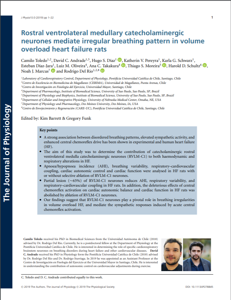
Rostral ventrolateral medullary catecholaminergic neurones mediate irregular breathing pattern in volume overload heart failure rats

Fecha
2019Autor
Andrade, David C. [Univ Mayor, Ctr Invest Fisiol Ejercicio, Santiago, Chile]
Toledo, Camilo; Díaz, Hugo S.; Pereyra, Katherin V.; Schwarz, Karla G.; Díaz-Jara, Esteban; Oliveira, Luiz M.; Takakura, Ana C.; Moreira, Thiago S.; Schultz, Harold D.; Marcus, Noah J.; Del Rio, Rodrigo
Ubicación geográfica
Notas
HERRAMIENTAS
Acceda a títulos restringidos
¿Cómo descargar?Resumen
Key points A strong association between disordered breathing patterns, elevated sympathetic activity, and enhanced central chemoreflex drive has been shown in experimental and human heart failure (HF). The aim of this study was to determine the contribution of catecholaminergic rostral ventrolateral medulla catecholaminergic neurones (RVLM-C1) to both haemodynamic and respiratory alterations in HF. Apnoea/hypopnoea incidence (AHI), breathing variability, respiratory-cardiovascular coupling, cardiac autonomic control and cardiac function were analysed in HF rats with or without selective ablation of RVLM-C1 neurones. Partial lesion (similar to 65%) of RVLM-C1 neurones reduces AHI, respiratory variability, and respiratory-cardiovascular coupling in HF rats. In addition, the deleterious effects of central chemoreflex activation on cardiac autonomic balance and cardiac function in HF rats was abolished by ablation of RVLM-C1 neurones. Our findings suggest that RVLM-C1 neurones play a pivotal role in breathing irregularities in volume overload HF, and mediate the sympathetic responses induced by acute central chemoreflex activation. Rostral ventrolateral medulla catecholaminergic neurones (RVLM-C1) modulate sympathetic outflow and breathing under normal conditions. Heart failure (HF) is characterized by chronic RVLM-C1 activation, increased sympathetic activity and irregular breathing patterns. Despite studies showing a relationship between RVLM-C1 and sympathetic activity in HF, no studies have addressed a potential contribution of RVLM-C1 neurones to irregular breathing in this context. Thus, the aim of this study was to determine the contribution of RVLM-C1 neurones to irregular breathing patterns in HF. Sprague-Dawley rats underwent surgery to induce volume overload HF. Anti-dopamine beta-hydroxylase-saporin toxin (D beta H-SAP) was used to selectively lesion RVLM-C1 neurones. At 8 weeks post-HF induction, breathing pattern, blood pressures (BP), respiratory-cardiovascular coupling (RCC), central chemoreflex function, cardiac autonomic control and cardiac function were studied. Reduction (similar to 65%) of RVLM-C1 neurones resulted in attenuation of irregular breathing, decreased apnoea-hypopnoea incidence (11.1 +/- 2.9 vs. 6.5 +/- 2.5 events h(-1); HF+Veh vs. HF+D beta H-SAP; P < 0.05) and improved cardiac autonomic control in HF rats. Pathological RCC was observed in HF rats (peak coherence >0.5 between breathing and cardiovascular signals) and was attenuated by D beta H-SAP treatment (coherence: 0.74 +/- 0.12 vs. 0.54 +/- 0.10, HF+Veh vs. HF+D beta H-SAP rats; P < 0.05). Central chemoreflex activation had deleterious effects on cardiac function and cardiac autonomic control in HF rats that were abolished by lesion of RVLM-C1 neurones. Our findings reveal that RVLM-C1 neurones play a major role in irregular breathing patterns observed in volume overload HF and highlight their contribution to cardiac dysautonomia and deterioration of cardiac function during chemoreflex activation.
Coleccion/es a la/s que pertenece:
Si usted es autor(a) de este documento y NO desea que su publicación tenga acceso público en este repositorio, por favor complete el formulario aquí.